Development and Optimization of Delivery, Imaging, and Collection Methods for Ocular Intravitreal Implants
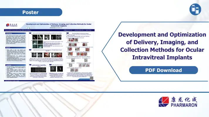
Poster Authors:
Glenwood Gum and Sandeep Kumar
Pharmaron (US) Lab Services LLC, San Diego, 92010, CA, United States
Ocular Intravitreal implants deliver drugs directly to the retina through controlled and sustained release. Getting implants placed safely requires precise surgical technique, imaging, and tissue collection to ensure correct positioning and understood degradation over time. Pharmaron’s ARVO 2026 poster presents optimized methods for implant delivery, real-time imaging and downstream tissue analysis across multiple species.
Intravitreal Implant Optimization
Poor IVT implant placement can lead to many complications that compromise study outcomes and delay programs. An additional layer of complications arise from different species. All species can have unique outcomes and without species specific procedural guidance, teams risk inconsistent implant positioning and unreliable data.
Poster Overview
This poster covers development & design across three areas essential to ocular implant optimization:
- Sclerotomy site selection
- Injection technique
- Multi-implant spacing
- in vivo imaging for Implant Monitoring
Capabilities
In order to track implant biodegradation over time, Pharmaron employed infrared (IR) imaging using Heidelberg Spectralis systems with both 55° and 102° lens configurations. Each lens serves a different purpose. The wider field lens confirms implant position. The higher magnification lens supports precise measurement of implant dimensions for quantifying erosion.
Fundus imaging was also used immediately after delivery to verify placement before animals recovered.
Tissue Collection for Downstream Analysis
Two dissection methods were developed for collecting ocular tissues after study completion:
- Fresh dissection
- Frozen dissection
Both approaches allow targeted sampling by tissue compartment and quadrant, with unique benefits specific to each study.
References:
- FDA Guidance: Nonclinical Safety Evaluation of Reformulated Drug Products and Products Intended for Administration by an Alternate Route
- NIH National Eye Institute
- Heidelberg Engineering Spectralis Platform
Download the Poster
This poster is helpful for researching pursuing intraocular sustained release implants, including gene therapy and small molecule programs that use IVT implant delivery. Download the full study to review species specific sclerotomy parameters, injection angle comparisons, IR imaging configurations and step-by-step dissection protocols for ocular tissue collection.
