Clinically Relevant Rabbit Models of Retinoblastoma and Choroidal Melanoma for Preclinical Evaluation of Ocular Oncology Therapies
Poster Authors:
Sandeep Kumar, Xiaohui Zhang; Sudan Puri; Matthew Lyulkin; Manindra Singh; Glenwood G. Gum
Pharmaron (US) Lab Services LLC, San Diego, 92010, CA, United States
Preclinical Ocular Oncology Models for Tumor Therapies
Preclinical ocular oncology models are essential for testing new treatments against eye cancers like retinoblastoma and choroidal melanoma. Without models that mimic how certain tumors grow and spread within the eye, it is difficult to predict the clinical success.
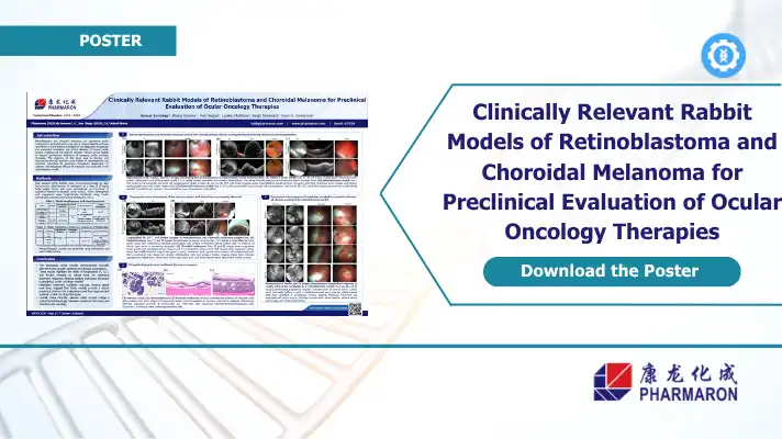
This Pharmaron poster, presented at ARVO 2026, details the development and characterization of rabbit models designed to replicate human ocular tumors. This study includes a validation method using melphalan in a retinoblastoma model.
Capabilities
Why Clinically Relevant Ocular Models Matter
Retinoblastoma and choroidal melanoma are aggressive cancers that carry a high risk of vitreous seeding and local recurrence, making treatment harder with an increased chance of relapse.
Many existing preclinical models don’t capture the full anatomical complexity of human ocular tumors. This gap limits the evaluation of how therapies interact with real tissue structures and disease progression patterns. Models that closely mirror clinical disease help researchers assess treatment response with greater confidence before moving into clinical trials.
What This Poster Covers
The poster presents rabbit models of retinoblastoma and choroidal melanoma developed at Pharmaron’s San Diego facility.
Key areas include:
- Model development using intraocular cell transplantation in immunocompromised New Zealand White rabbits
- Longitudinal monitoring of tumor growth using infrared imaging (IR), optical coherence tomography (OCT) and fundus photography
- Histopathology of tumor characterization
- Preclinical ocular evaluation to assess antitumor response and vitreous seed reduction
Imaging and Monitoring
The models highlighted in this study used a combination of IR, OCT, and fundus imaging at multiple time points in order to monitor tumor growth patterns, vitreous involvement, and treatment response.
Relevance for Ocular Oncology Drug Development
By reproducing behavior like vitreous seeding and progressive choroidal tumor expansion, the models offer a more realistic platform for evaluating regional and systemic therapies.
Access to validated animal models with multimodal imaging endpoints can support stronger IND-enabling data packages and better-informed study designs.
Download the Poster
Download the full ARVO 2026 poster to learn more.
References:
