Tissue Distribution with QWBA and mARG
Tissue Distribution with QWBA and mARG Services
Pharmaron’s quantitative whole-body autoradiography (QWBA) capabilities enable rapid quantitation of tissue distribution in a variety of animal models. More detailed examination can also be conducted at the intra-organ and cellular level using microautoradiography (mARG). This is complemented by traditional tissue dissection, analysis (“cut and burn”) and histology techniques where appropriate. Both techniques can be combined with an integrated service to provide 14C and 3H radiolabels for use in these programs. These approaches can be employed to study small molecules, large molecules, polymers, conjugates and delivery devices.
Related Downloads
Learn more about Pharmaron’s Tissue Imaging Services Using Radiolabelled Compounds.
Capabilities
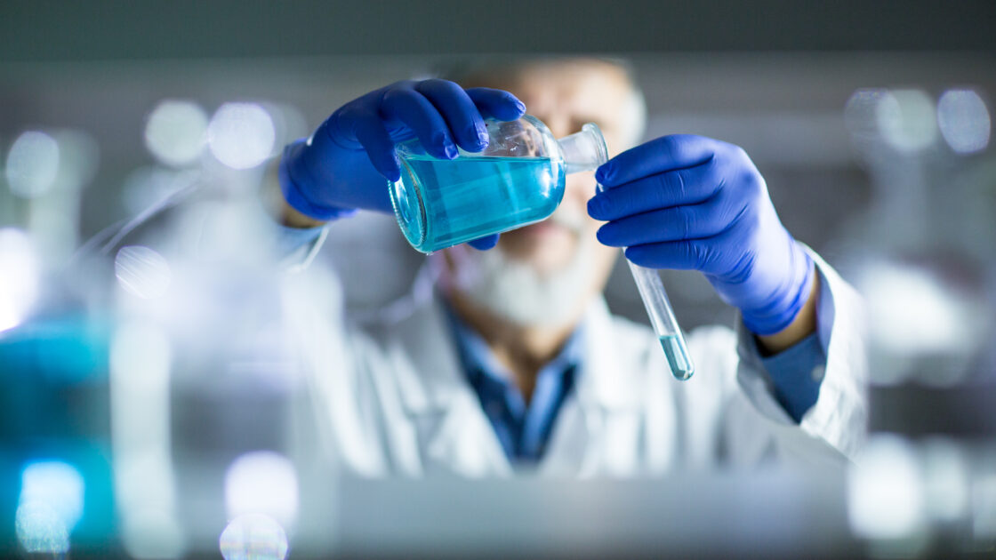
Radiolabelled Chemical Synthesis
- Bespoke radiosynthesis of 14C and 3H small molecules
- Radiolabelling of peptides, proteins, antibody-drug conjugates (ADCs), polymers and other constructs
- Radiolabelled oligonucleotide synthesizer
- Rapid covalent radiolabelling techniques with 3H/14C to “RadioTag” macromolecules
0
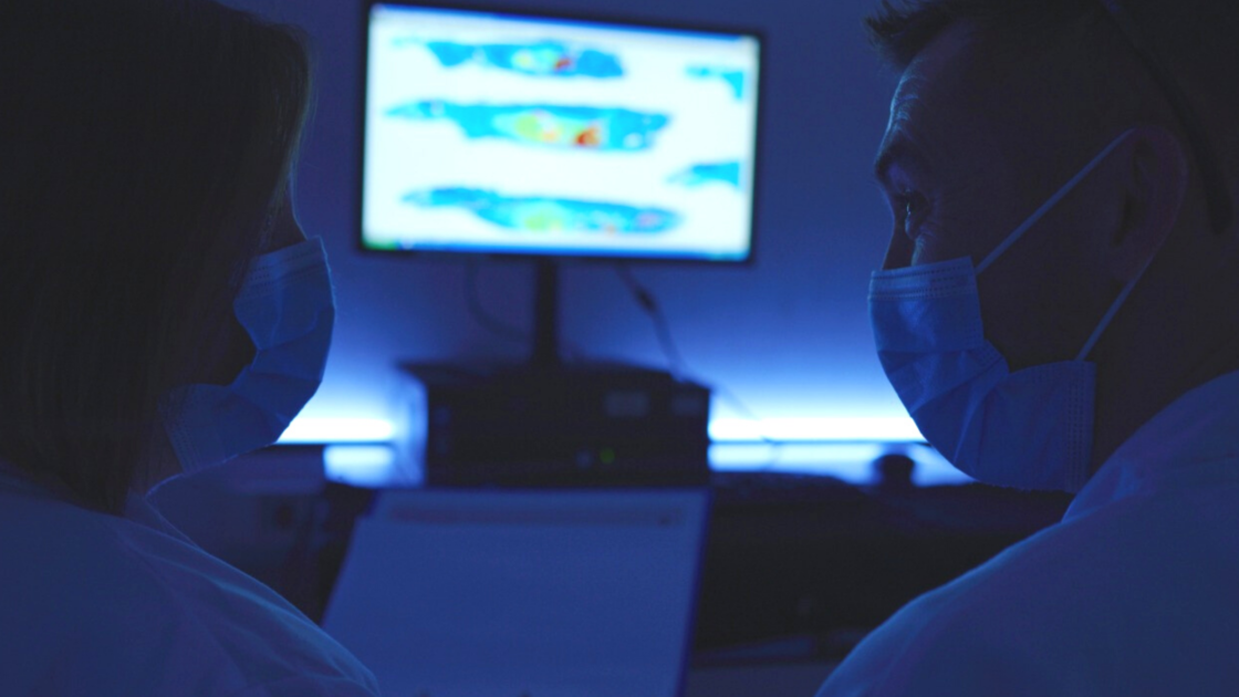
Quantitative Whole Body Autoradiography (QWBA)
- Distribution of drug-related material in blood and tissues
- Target organ engagement for drug-related material (e.g. CNS, kidney, liver)
- Identify sites of accumulation or disposition
- Assess melanin-binding in pigmented tissues
- Fetal exposure and milk secretion to support teratology studies
- Human dosimetry calculations and dosimetry reports for clinical metabolism studies
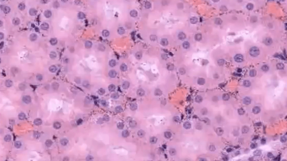
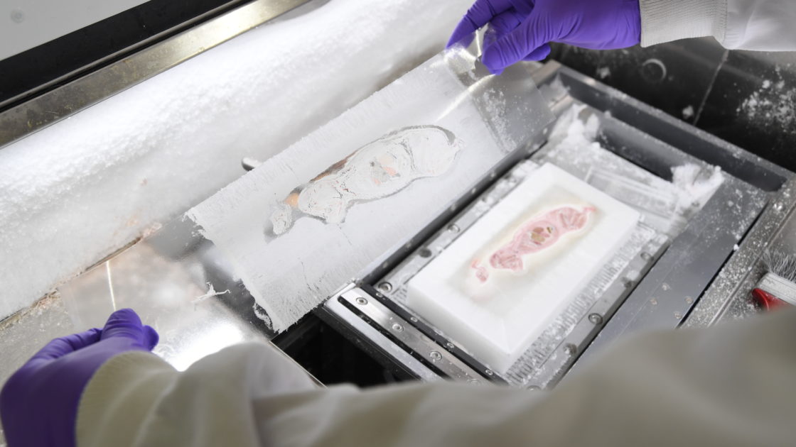
Microautoradiography (mARG)
- High resolution for determining intra-organ distribution
- Intracellular distribution in specific cell types
- Identification of binding sites within organs and cells
- Receptor occupancy studies and immunohistochemistry
0
Novel Real-time Digital Autoradiography
- Fast turnaround tritiation and tissue imaging (3H) to support late-stage lead optimization or candidate selection
- Concomitant differential tissue distribution for dual 3H/14C radiolabelled compounds – both 3H and 14C radiolabels can be quantified simultaneously in the same tissue slices
- Autoradiographs are available in hours rather than days
0
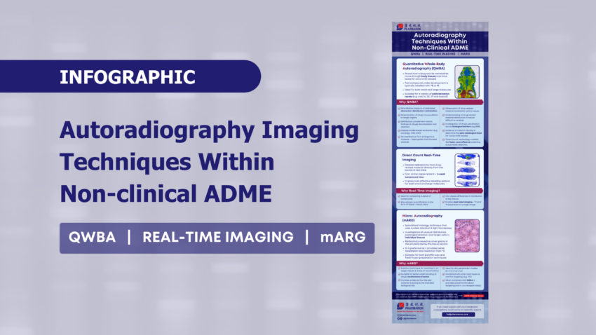
Download infographic to learn more about the Autoradiography Imaging Techniques Within Non-Clinical ADME.