in vitro Disease Biology

in vitro Disease Biology Services
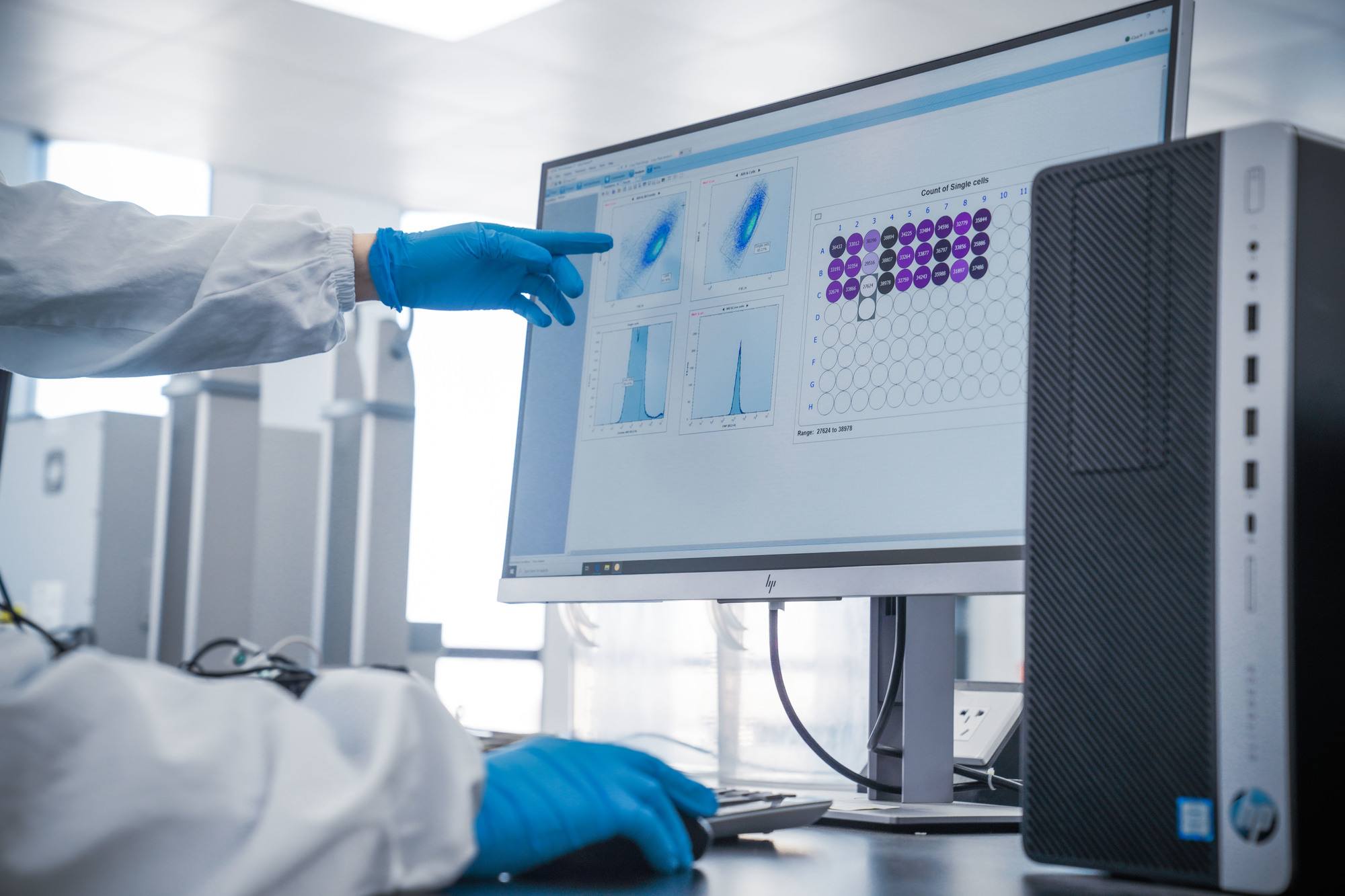
Our in vitro disease biology team works closely with our partners to evaluate targets, design flexible work flows, and develop and validate assays to meet project specific goals. Pharmaron has strong assay development expertise in key therapeutic areas, including oncology, immunology, IO, metabolic disease, cardiovascular disease, CNS and pain.
Target-based screening platforms and pathways-centric approaches can be applied to multiple diseases and therapeutic areas.
Capabilities
Oncology
- Target validation
- Drug-resistant cell line development and characterization
- Cell viability
- Long-term culture, 2D, 3D
- 3D spheroid assays (viability/Western blot/qPCR)
- Apoptosis assays
- Cell cycle analysis
- DNA damage (rH2Ax, gene pathways)
- Angiogenesis
- Migration/invasion assay; translocation assay

Immunology
- T cell activation/proliferation
- PBMC assays
- Whole-blood assays
- Toll-like receptor
- cGAS-STING pathway
- JAK/STAT pathway
- IRF pathway
- NF-KB pathway
- NF-AT pathway
- PD-1/PD-L1 blockage
- IDO/TDO/Adenosine receptors
- Stress pathway exploration
Cardiovascular and Metabolic Diseases
- GSIS (Intact islet/pancreatic beta cell)
- 3T3-L1 differentiation
- Fatty acid synthesis assay (C-14)
- Glucose uptake on different cell models
- Ephys-based ion channel assays
- Ephys-recording from primary culture
- iPS-induced cardiomyocyte
- Multiple-electrode array recording of cardiomyocyte
CNS
- A beta aggregation
- High content-based reading from primary cultured neuron
- Chemical-induced neurotoxicity, neurite degeneration model
- Ephys-based ion channel assays
- Ephys-recording from primary culture
- Mechanical induction of neurotoxicity, neurite degeneration model
- Neurite outgrowth